/ H2A / Variant: H2A.X
Description
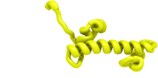
H2A.X most notably is involved in DNA damage response, marking the double strand DNA breaks with its phosphorylated form ɣ-H2A.X. It is also more broadly implicated in chromatin remodeling, found at collapsed replication forks and heterochromatin. It is the only non-monophyletic variant in H2A and has the charactristic SQE/DΦ motif at C-terminus (SQAY in Drosophila), where Φ-represents a hydrophobic residue (usually Tyr in mammals), and S is phosphorylation site. Nematodes are the only known species that lack H2A.X. In yeast H2A.X function is fulfilled by conventional H2A, while in Drosophila melanogaster by H2A.Z.Alternate names: member X
Features
Below, you can explore the features of H2A.X from Homo sapiens, if available and how it compares to the canonical histones of the same type (first row). Canonical histone is shown in the first row, the names and descriptions of each feature can be found underneath. To explore variants from other species, please browse our curated sequences, automatically extracted sequences, or by taxonomy.
Keys: red - identical residues, blue - different residues (if more than one sequence).
Variant features
mSph
Mouse Serine phosphorylation site
Kac
Human Lysine acetylation site
Kbio
Mouse Lysine biotinylation site
mKac
Mouse Lysine acetylation site
Kub
Human, rat and mouse Lysine ubiquitination site
Sph
Human Serine phosphorylation site
H2A.X-motif
SQE/DΦ motif at C-terminus; Φ-hydrophobic residues, usually Tyr in mammals
p
C-terminal serine can be phosphorylated in H2A.X-containing nucleosomes at sites of DNA double strand breaks.
Yph
Human and mouse Tyrosine phosphorylation site
General histone type features
alpha1ext
Alpha1-extension helix
alpha1
Alpha1-helix, first helix of histone fold
loopL1
L1 loop, connecting first and second helices of histone fold. Part of L1L2 DNA binding site formed by H2A and H2B at SHL ±3.5.
R1
Minor groove arginine at L1L2 DNA binding site, SHL ±3.5
beta1
Beta-strand in L1L2 DNA binding site
alpha2
Alpha2-helix, second helix of histone fold
ap
Acidic patch residues
loopL2
L2 loop, connecting second and third helices of histone fold. Part of L1L2 DNA binding site formed by H2A and H2B at SHL ±5.5.
R2
Minor groove arginine at L1L2 DNA binding site, SHL ±5.5
beta2
Beta-strand in L1L2 DNA binding site
alpha3
Alpha3-helix, third helix of histone fold
Docking domain
Docking domain locking H2A-H2B dimer on H3-H4 tetramer surface
alpha3ext
Alpha3-extension helix
beta3
Beta-strand between H2A and H4
References
- Talbert PB, Ahmad K, et al. "A unified phylogeny-based nomenclature for histone variants." Epigenetics Chromatin, 2012. PMID: 22650316
- Shaytan AK, Landsman D, et al. "Nucleosome adaptability conferred by sequence and structural variations in histone H2A-H2B dimers." Curr Opin Struct Biol, 2015. PMID: 25731851
- Ismail IH and Hendzel MJ. "The gamma-H2A.X: is it just a surrogate marker of double-strand breaks or much more?" Environ Mol Mutagen, 2008. PMID: 18095327
- Millar CB. "Organizing the genome with H2A histone variants." Biochem J, 2013. PMID: 23301656
A set of manually selected and validated histone sequences is listed in the table. Click on an entry in the table to update the annotated sequence preview: a variant will be compared with the canonical histone from the same species (if available).
Alternatively, tick mark the sequences and use toolbar to view MSA, export or add to basket. Use search or filters to find particular entries.
Keys: red - identical residues, blue - different residues (if more than one sequence). For feature legend see summary tab.
Sequence preview and annotation... LOADING
Min Score
Max Score
168.3
239.7
You selected: root
Note: variant classification might be ambigous between very similar variants. Classification scores against all variant models are available via advanced menu.
Features characteristic for a given histone type/variant are marked below the consensus sequence. For feature description see summary tabs of the corresponding variants pages.
Keys: red - 80% identical, blue - 50% identical columns. X-ambigous positions in consensus sequence.